
COMMON EYE DISORDERS | EDUCATION CENTER

OCULAR ANATOMY | EDUCATION CENTER
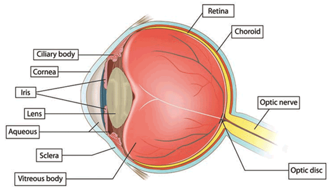
The average width of the adult human eyeball is approximately 24 mm wide. A tough white covering called the sclera protects the eye. Part of the white sclera can be seen in the front of the eye. A clear, delicate membrane called the conjunctiva covers the sclera.
At the front of the eye is the cornea. The cornea is the clear part of the eye’s protective covering. It allows light to enter the eye. The iris is the colored part of the eye that contracts and expands so the pupil can let just the right amount of light into the eye. The light is directed by the pupil to the lens. The lens focuses the light onto the retina (lining the back of the eye). Nerve fibers in the retina carry images to the brain through the optic nerve.
The front part of the eye is filled with a clear fluid called intraocular fluid or aqueous humor, made by the ciliary body. The fluid flows out through the pupil. It is then absorbed into the bloodstream through the eye’s drainage system.
This drainage system is a meshwork of drainage canals around the outer edge of the iris. Proper drainage helps keep eye pressure at a normal level. The production, flow, and drainage of this fluid is an active continuous process that is needed for the health of the eye.
The inner pressure of the eye (intraocular pressure or IOP) depends upon the amount of fluid in the eye. If your eye’s drainage system is working properly then fluid can drain out and prevent a buildup. Likewise, if your eye’s fluid system is working properly, then the right amount of fluid will be produced for a healthy eye. Your IOP can vary at different times of the day, but it normally stays within a range that the eye can handle.
You have millions of nerve fibers that run from your retina to the optic nerve. These fibers meet at the optic disc. Visual information is transferred from the optic nerve to the visual cortex in the brain.
The visual cortex of the brain is that part of the cerebral cortex which processes visual information. It is located in the occipital lobe. Visual nerves run straight from the eye to the primary visual cortex to the visual association cortex.
Source: American Optometric Association (https://www.aoa.org/)

The Visual Cortex occupies the posterior Occipital Lobe and is connected to the eyes via the optic nerve.